
|
Lehigh bursts onto the blockchain scene. | READ MORE >> |
Computer Science and Engineering is at the core of the information age. The explosion of data brought about in the last two decades by the revolution in digital technology and the Internet has reshaped our world, and yet its really just begun.
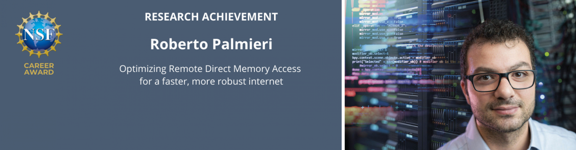
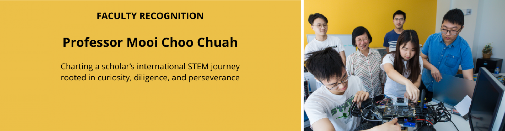
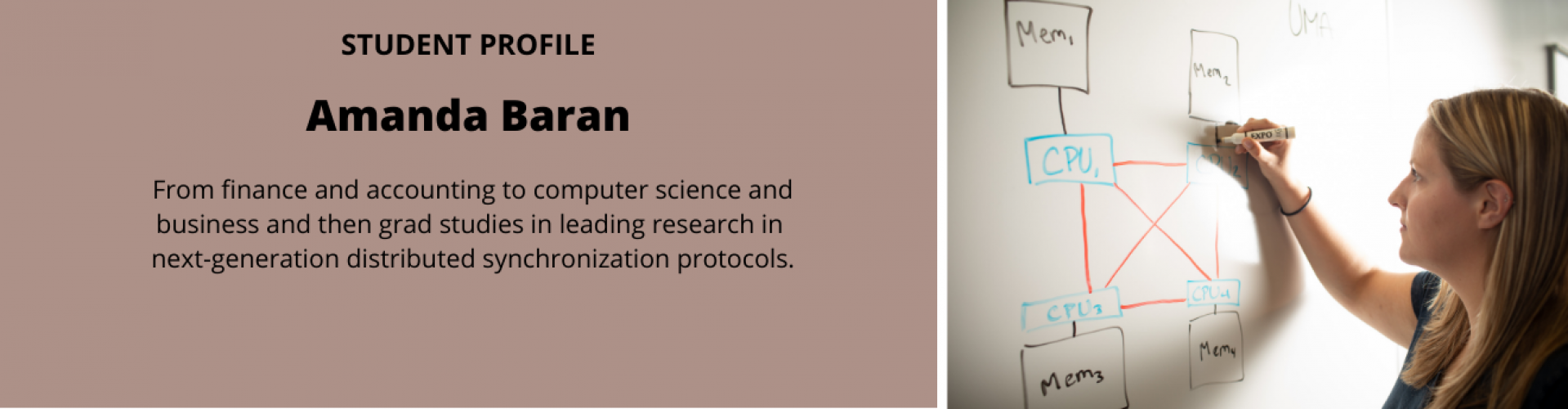
As leaders in research, CSE faculty members work with students of all levels, our internationally-recognized faculty conduct groundbreaking research in artificial intelligence, bioinformatics, data mining, robotics, software security, computer networking, software systems, biomedical image processing, computer vision, mobile healthcare, and the Web. Our faculty have research funded by competitive sources including NSF, DARPA, NIH, and other federal and state agencies, as well as leading companies in the field.
We encourage you to browse our website, visit individual faculty pages, or contact us to learn about our dynamic research and graduate and undergraduate educational programs. | READ MORE >>
Department News
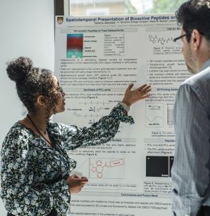
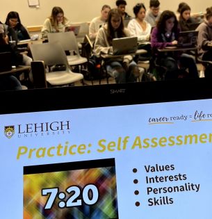
Undergraduate research takes the spotlight on April 19

Students looking for an elective? See what ChBE has planned for fall